
Kinase Knowledgebase · contact maps
What the ligand is actually doing
Every compound in the Kinase Knowledgebase that matches a PDB kinase ligand exactly by canonical SMILES, and that KKB has measured against that same target, gets a contact map with typed, directional interactions rather than a list of close atoms. Nothing here is predicted. Every number is geometry computed from the deposited coordinates.
PIK3CA with ligand FE5, PDB 6GVF
What a kinase ligand is actually doing, drawn from measured geometry: hydrogen bonds with real donor to hydrogen to acceptor angles, halogen bonds, salt bridges, pi stacking, cation to pi and metal coordination, measured from deposited PDB coordinates and paired with Kinase Knowledgebase activity for the same compound against the same target.
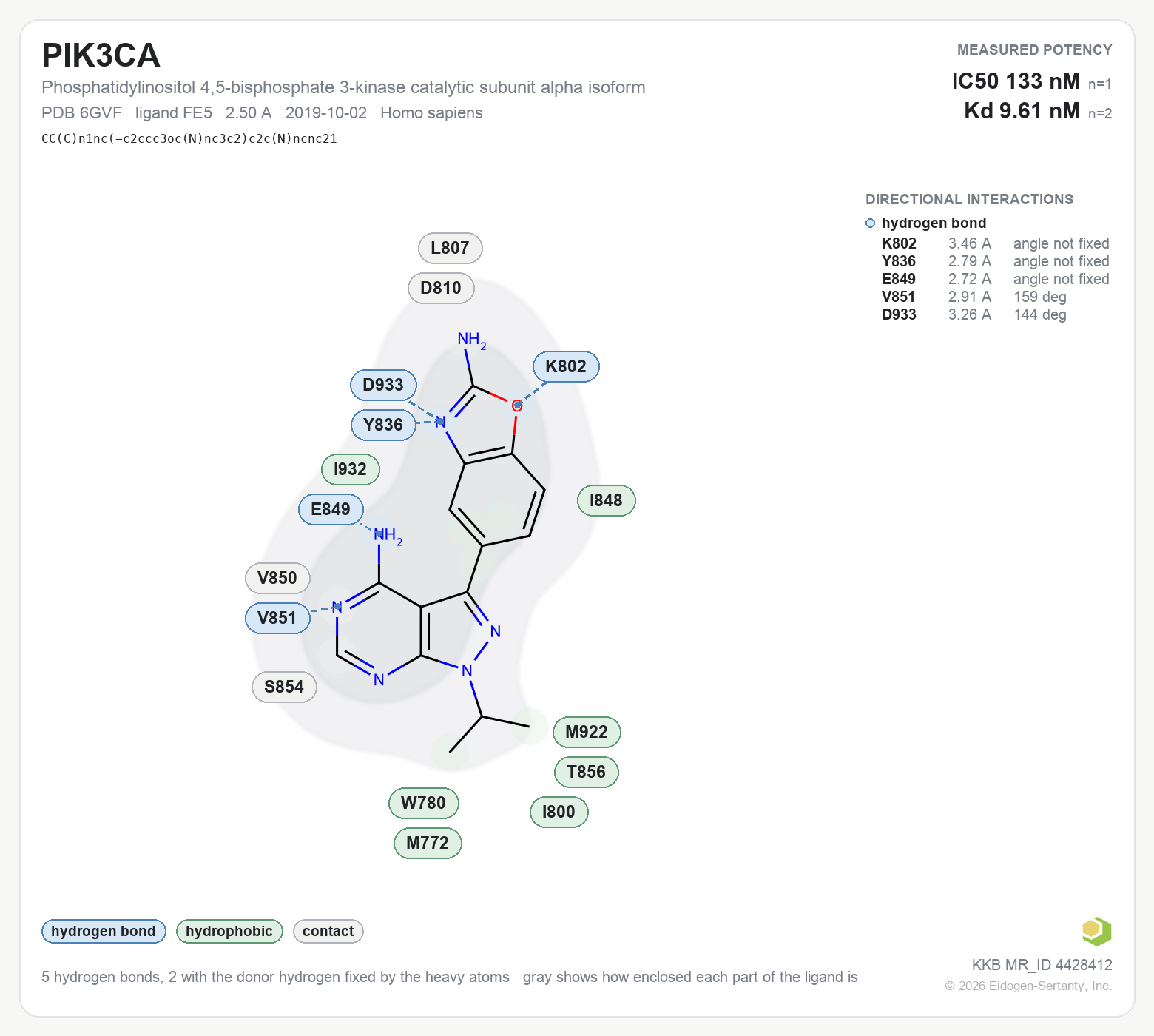
The rest of this page is how that figure was computed. It applies to every one of them.
What is measured
All distances are between heavy atoms as deposited, first alternate location only, model 1 only. The cutoffs travel with every figure in its data record, so any number on a figure can be checked against the structure it came from. There is no model and no judgment anywhere in this: the interactions are computed by deterministic geometry, and no interaction package is used.
Hydrogen bond
Donor to acceptor within 3.5 A, hydrogen to acceptor within 2.7 A, a donor to hydrogen to acceptor angle of at least 120 degrees, and a sensible angle at both antecedent atoms.
Halogen bond
A carbon to halogen to acceptor angle of at least 140 degrees, within 0.4 A of the van der Waals sum. Needs no modeled hydrogen at all.
Salt bridge
Opposite formal charges within 4 A, at standard states for neutral pH. Histidine is not assumed charged.
Pi stacking
Ring centroids within 5.5 A. Parallel below 30 degrees between ring planes, edge to face above 60 degrees.
Cation to pi
A lysine or arginine cation within 6 A of a ring centroid and within 35 degrees of the ring normal.
Metal coordination
Magnesium, manganese, zinc and the rest within 3 A of a ligand nitrogen, oxygen or sulfur.
Hydrophobic
Carbon to carbon within 4.5 A, excluding carbons bonded to nitrogen, oxygen or fluorine, which are polarized.
Bridging water
A water is drawn only when its oxygen is within 3.5 A of a ligand nitrogen, oxygen or sulfur and independently within that distance of a protein nitrogen, oxygen or sulfur. A water touching one side is not drawn. These are waters that bridge the two.
Close contact, the leftover class
Each residue is colored by its strongest interaction. Gray means a heavy atom pair within 4.5 A where that residue makes nothing more specific: a close polar pair with no hydrogen to give, a carbon to oxygen or carbon to nitrogen approach, or a carbon to carbon approach where one carbon is polarized. It exists so a residue that genuinely lines the site is not dropped. It is a close contact, not a weak interaction.
Hydrogens
At these resolutions hydrogens are not observed, so none is ever read from the file. Where the heavy atoms fix a hydrogen's position, as they do for a backbone amide NH, an aromatic NH or a guanidinium, the real donor to hydrogen to acceptor angle is computed and reported.
Where the donor can turn freely, as a serine hydroxyl, a lysine ammonium or a water can, no orientation is invented. The panel says the angle is not fixed, and the contact is reported only as geometrically satisfiable, meaning some rotamer of that donor would give an acceptable angle. That is the strongest true statement the data supports.
A hydrogen bond on these figures is never called proven. It satisfies the stated geometry, and the figure says which of the two cases it is.
When the two records disagree on 3D form
A figure is drawn only when the compound with the measured activity is the same molecule as the ligand in the structure. Sometimes the two records describe that molecule with different precision, and where they do, the figure says so under the SMILES and rings the atoms involved.
- The structure is more specific. The entry shows one specific 3D form and the Kinase Knowledgebase does not record which form was tested, so the potency may be for a mixture. That is the limit of what a measured number can say about a drawn isomer.
- The activity record is more specific. The Kinase Knowledgebase records which form was tested and the structure entry does not.
- Double bond geometry. The two records name the marked double bond differently, cis in one and trans in the other.
A molecule with a genuinely inverted center is a different compound, and is never drawn.
The gray envelope
The gray shape behind each ligand is a count, per ligand atom, of protein heavy atoms within 6 A, smoothed onto the flat drawing. It shows how enclosed each part of the molecule is. It is a burial measure, not a cross section of the pocket.
Bound is not necessarily potent → Fourteen protein sites, every co-crystal ligand superposed into one frame with its measured potency. Within one pocket the values span up to six orders of magnitude. The kinase rows, CDK2, CHK1, JAK2 and BTK, rest on Kinase Knowledgebase data.Talk to us about the Kinase Knowledgebase
These maps sit on top of KKB structure activity data. We are happy to walk through the method, the coverage and what it is useful for.